Is There a Nonperfusion Threshold on OCT Angiography Associated With New Vessels Detected on Ultra-Wide-Field Imaging in Diabetic Retinopathy? Nonperfusion Area to Diagnose Diabetic Retinopathy
Hugo Le Boité, Alain Gaudric, Ali Erginay, Ramin Tadayoni, Aude Couturier
Abstract
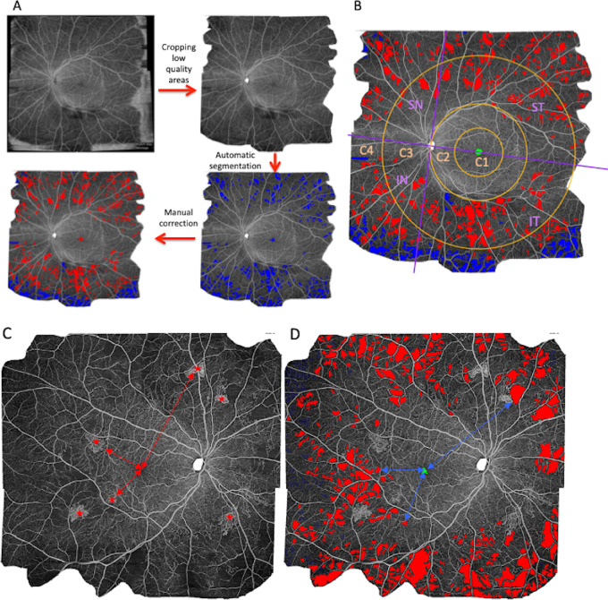
'Aude Couturier'] Purpose To determine whether the nonperfusion index (NPI) measured on widefield (WF) optical coherence tomography angiography (OCTA) could be used as an alternative method for the diagnosis of proliferative diabetic retinopathy (PDR) and to study the relationship between the NPI and the location of new vessels (NV) in eyes with PDR. Methods Fifty-one treatment-naïve eyes with either severe nonproliferative DR (NPDR) or PDR were imaged using ultra-wide-field imaging and wide-field OCTA. Results The NPI was significantly higher in eyes with PDR (18.94% vs. 7.51%; P < 0.01). Using the NPI on the whole image to assess PDR status, the area under the curve was 0.770, but the area under the curve increased when the NPI of the most peripheral circle was used (area under the curve of 0.792). Four eyes with PDR (17%) had NV outside the OCTA image field, and their mean NPI (6.15 %) did not differ from that measured in severe NPDR eyes (7.51%; P = 0.67) and was lower than in other eyes with PDR (21.49%; P = 0.023). The presence of NV in a sector was associated with a higher NPI in the same sector (29.2% vs. 6.0%; P < 10−15). Conclusions Although the NPI was significantly higher in eyes with PDR compared with severe NPDR eyes, its measurement on the whole wide-field OCTA image was not sensitive enough to replace the detection of NV for the diagnosis of PDR. Translational Relevance Because the presence of new vessels was related to the local nonperfusion index in the same sector, the assessment of nonperfusion outside the optical coherence tomography angiography field is important in diabetic retinopathy.
§ The Valyu brief
Reading the full paper and taking notes. This takes a few seconds…
§ Ask this paper
Ask a question about this paper
Valyu reads the full text and answers from what the paper actually says.
Searching the other archives…