Follicle-like niches outside the cortex? 3D phase-contrast µCT revealed medullary B cell nodules in mucosa-draining lymph nodes
Paul Schütz, Florian L. Schwarzenberg, Lennert J. Weber, Jörg U. Hammel, Bente Siebels, Paula Nissen, Nenya Leising, Katja J. Jarick, Bernd Walkenfort, Sarah C. Irvine, Jasmin Bartl, Julia Herzen, Christian Lohr, Clemens Wülfing, Stephan Henne
Abstract
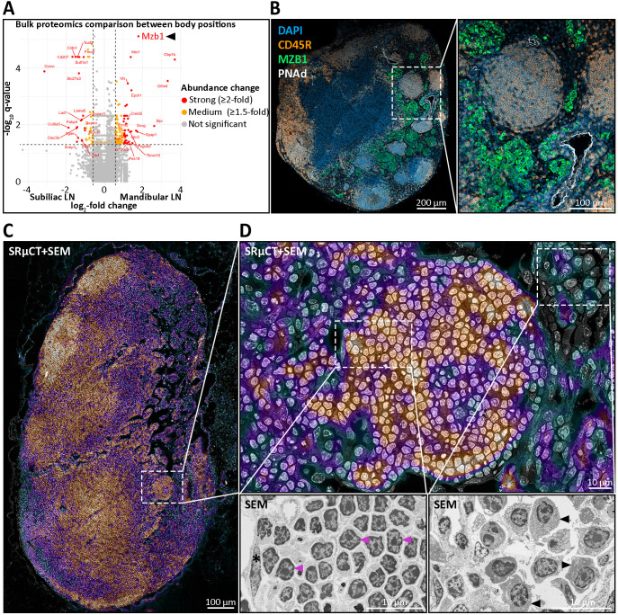
This study identifies and characterizes previously unrecognized medullary B cell niches within murine mucosa-draining lymph nodes (LNs), challenging the conventional understanding of LN architecture. Utilizing advanced imaging techniques, including synchrotron radiation-based phase-contrast micro-computed tomography (SRµCT), correlated high-resolution electron microscopy and immunohistochemistry (IHC), we revealed spherical to ovoid structures termed nodules, being distinct lymphoid compartments consistently localized in the medullary region of mandibular and other mucosa-draining LNs. These nodules were primarily composed of unswitched, non-proliferative CD45R+ B cells expressing IgD and IgM, lacking germinal center features or typical activation markers. They were seamlessly integrated into the medullary architecture, surrounded by LYVE-1+ lymphatic endothelial cells, and situated in close proximity to medullary high endothelial venules (HEVs), revealed by PNAd staining. Under steady-state conditions, this has not been previously observed in the medullary compartment of LNs but is likely facilitating nodule-like B cell aggregation in mucosa-draining LNs due to sustained low level antigenic stimulation common in mucosal environments and is underpinned by proteomics. Additionally, the nodules displayed a capillary network that closely resembles the vascularization seen in conventional B cell follicles revealed by SRµCT. Nodule formation occured between two and four weeks postnatally, thus emerging later than B cell follicles, and their abundance showed a tendency to increase with age. Functionally, these nodules appear to represent a quiescent B cell niche, potentially supporting B cell homeostasis, tolerance, or memory-like readiness, and are distinct from pathological hyperplasias. Their preservation in aged LNs, coupled with the absence of lipomatosis, suggests a role in maintaining structural integrity and immune readiness through persistent B cell-stromal interactions.
§ The Valyu brief
Reading the full paper and taking notes. This takes a few seconds…
§ Ask this paper
Ask a question about this paper
Valyu reads the full text and answers from what the paper actually says.
Searching the other archives…