Segmentation of Choroidal Boundary in Enhanced Depth Imaging OCTs Using a Multiresolution Texture Based Modeling in Graph Cuts
Hajar Danesh, Raheleh Kafieh, Hossein Rabbani, Fedra Hajizadeh
Abstract
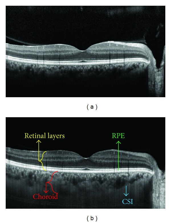
The introduction of enhanced depth imaging optical coherence tomography (EDI-OCT) has provided the advantage of in vivo cross-sectional imaging of the choroid, similar to the retina, with standard commercially available spectral domain (SD) OCT machines. A texture-based algorithm is introduced in this paper for fully automatic segmentation of choroidal images obtained from an EDI system of Heidelberg 3D OCT Spectralis. Dynamic programming is utilized to determine the location of the retinal pigment epithelium (RPE). Bruch's membrane (BM) (the blood-retina barrier which separates the RPE cells of the retina from the choroid) can be segmented by searching for the pixels with the biggest gradient value below the RPE. Furthermore, a novel method is proposed to segment the choroid-sclera interface (CSI), which employs the wavelet based features to construct a Gaussian mixture model (GMM). The model is then used in a graph cut for segmentation of the choroidal boundary. The proposed algorithm is tested on 100 EDI OCTs and is compared with manual segmentation. The results showed an unsigned error of 2.48 ± 0.32 pixels for BM extraction and 9.79 ± 3.29 pixels for choroid detection. It implies significant improvement of the proposed method over other approaches like k-means and graph cut methods.
§ The Valyu brief
Reading the full paper and taking notes. This takes a few seconds…
§ Ask this paper
Ask a question about this paper
Valyu reads the full text and answers from what the paper actually says.
Searching the other archives…