Increase in Brain Volume After Aneurysmal Subarachnoid Hemorrhage Leads to Unfavorable Outcome: A Retrospective Study Quantified by CT Scan
Bin Qin, Yi Xiang, Jianfeng Zheng, Rui Xu, Zongduo Guo, Chongjie Cheng, Li Jiang, Yue Wu, Xiaochuan Sun, Zhijian Huang
Abstract
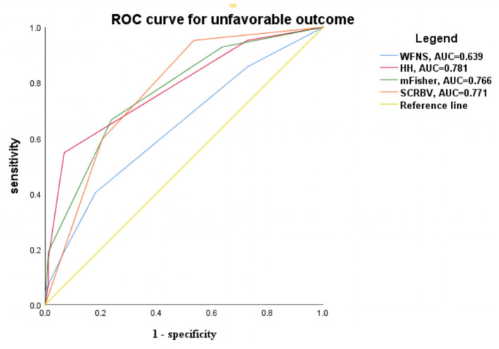
'Chongjie Cheng' 'Li Jiang' 'Yue Wu' 'Xiaochuan Sun' 'Zhijian Huang'] Background and Purpose: Primary brain swelling occurs in aneurysmal subarachnoid hemorrhage (aSAH) patients. The absence of a dynamic quantitative method restricts further study of primary brain swelling. This study compared differences in the change rate of brain volume (CRBV) between patients with and without primary brain swelling in the early stage of aSAH. Moreover, the relationship between CRBV and clinical outcomes was evaluated. Methods: Patients hospitalized within 24 h after aSAH were included in this retrospective study. Utilizing a qualitative standard established before the study to recognize primary brain swelling through brain CT after aSAH, clinical outcomes after 3 months of SAH were evaluated with a modified Rankin scale (mRS). The brain volume (BV) of each patient was calculated with a semiautomatic threshold algorithm of 3D-slicer, and the change in brain volume (CIBV) was obtained by subtracting the two extreme values (CIBV = BVmax - BVmin). The CRBV was obtained by CIBV/BVmin × 100%. The CRBV values that predicted unfavorable prognoses were estimated. Results: In total, 130 subjects were enrolled in the study. The mean CRBV in the non-swelling group and swelling group were 4.37% (±4.77) and 11.87% (±6.84), respectively (p < 0.05). CRBV was positively correlated with the length of hospital stay, blood in the ambient cistern, blood in the lateral ventricle, and lateral ventricular volume (Spearman ρ = 0.334; p < 0.001; Pearson ρ = 0.269, p = 0.002; Pearson ρ = 0.278, p = 0.001; Pearson ρ = 0.233, p = 0.008, respectively). Analysis of variance showed significant differences in CIBV, CRBV, blood in the ambient cistern, blood in the lateral ventricle, and lateral ventricular volume among varying modified Fisher scale (mFisher), with higher admission mFisher scale, indicating larger values of these variables. After adjusting for risk factors, the model showed that for every 1% increase in the CRBV, the probability of poor clinical prognosis increased by a factor of 1.236 (95% CI = 1.056-1.446).
§ The Valyu brief
Reading the full paper and taking notes. This takes a few seconds…
§ Ask this paper
Ask a question about this paper
Valyu reads the full text and answers from what the paper actually says.
Searching the other archives…