Imaging of lumpectomy surface with large field-of-view confocal laser scanning microscope for intraoperative margin assessment - POLARHIS study
Mariana-Felicia Sandor, Beatrice Schwalbach, Viktoria Hofmann, Simona-Elena Istrate, Zlatna Schuller, Elena Ionescu, Sara Heimann, Moira Ragazzi, Michael P. Lux
Abstract
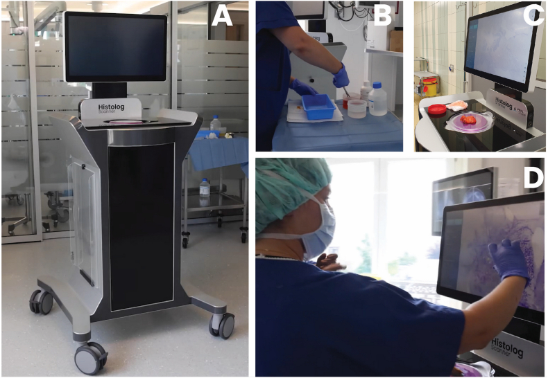
'Simona-Elena Istrate' 'Zlatna Schuller' 'Elena Ionescu' 'Sara Heimann' 'Moira Ragazzi' 'Michael P. Lux'] Introduction Breast-conserving surgery (BCS) in case of breast cancer and/or in-situ-carcinoma lesions (DCIS) intends to completely remove breast cancer while saving healthy tissue as much as possible to achieve better aesthetic and psychological outcomes for the patient. Such modality should result in postoperative tumor-free margins of the surgical resection in order to carry on with the next therapeutical steps of the patient care. However, 10-40% of patients undergo more than one procedure to achieve acceptable cancer-negative margins. A 2nd operation or further operation (re-operation) has physical, psychological, and economic consequences. It also delays the administration of adjuvant therapy, and has been associated with an elevated risk of local and distant disease relapse. In addition, a high re-operation rate can have significant economic effects - both for the service provider and for the payer. A more efficient intraoperative assessment of the margin may address these issues. Recently, a large field-of-view confocal laser scanning microscope designed to allow real-time intraoperative margin assessment has arrived on the market - the Histolog Scanner. In this paper, we present the first evaluation of lumpectomy margins assessment with this new device. Materials and methods 40 consecutive patients undergoing BCS with invasive and/or DCIS were included. The whole surface of the surgical specimens was imaged right after the operation using the Histolog Scanner (HLS). The assessment of all the specimen margins was performed intraoperatively according to the standard-of-care of the center which consists of combined ultrasound (IOUS) and/or conventional specimen radiography (CSR), and gross surgical inspection. Margin assessment on HLS images was blindly performed after the surgery by 5 surgeons and one pathologist. The capabilities to correctly determine margin status in HLS images was compared to the final histopathological assessment.
§ The Valyu brief
Reading the full paper and taking notes. This takes a few seconds…
§ Ask this paper
Ask a question about this paper
Valyu reads the full text and answers from what the paper actually says.
Searching the other archives…